Figure 4.
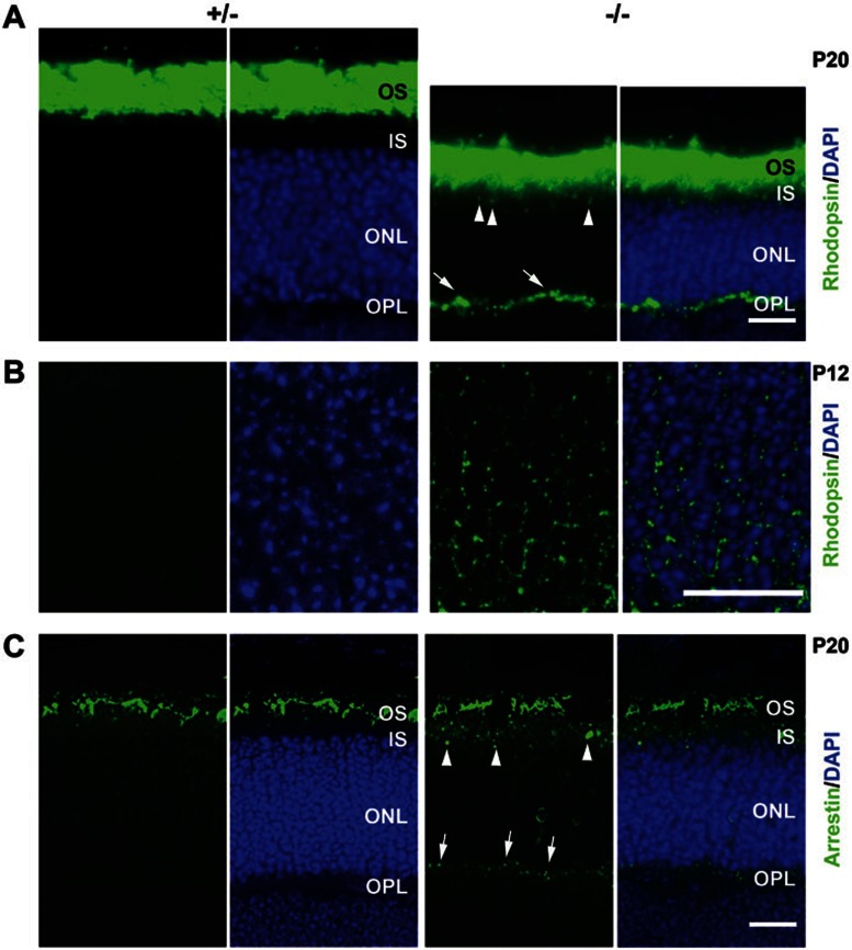
Mislocalization of OS proteins in Dlic1−/− photoreceptor cells. (A) Immunofluorescent staining of cryosections of P20 Dlic1+/− and Dlic1−/− retinas with anti-rhodopsin reveals the mislocalization of rhodopsin in Dlic1−/− photoreceptor cells. (B) Higher magnification of P12 mouse retinas immunofluorescent -stained with anti-rhodopsin. Perinuclear mislocalized rhodopsin is only shown in the Dlic1−/− retina. (C) Immunofluorescent-stained cryosections of P20 Dlic1+/− and Dlic1−/− retinas with anti-arrestin. Arrows and arrowheads in A and C indicate the mislocalization of rhodopsin or arrestin in the OPL and the IS, respectively. Cell nuclei were stained with DAPI. Images are representative retina sections from at least three mice per group. Bar = 20 μm.

