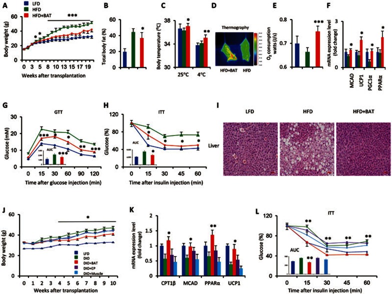
Figure 1.
BAT transplantation prevents or reverses HFD-induced obesity. (A–I) To test the ability of BAT to prevent the development of obesity, HFD feeding was initiated immediately after BAT transplantation. BAT was dissected from strain-, sex- and age-matched donor mice, and was subcutaneously transplanted into the dorsal interscapular region of recipient mice (6-week-old males). The results showed that BAT transplantation (A) reduced the HFD-induced weight gain; (B) decreased the total fat mass, as determined by computerized tomography performed 17 weeks post transplantation (wpt); (C) increased the core body temperature under thermoneutral or cold conditions, assessed 19 wpt; (D) significantly increased the heat production during cold challenge, as indicated by thermographic imaging performed 12 wpt; (E) increased oxygen consumption, as determined by a TSE lab master system performed 11 wpt; (F) increased the mRNA expression of fatty acid oxidation-related genes in BAT, as determined by qPCR at the end of the study; (G–H) improved the HFD-induced insulin resistance, as determined by (G) GTT (inner graph, area under the curve (AUC) for GTT) performed 12 wpt and (H) ITT (inner graph, AUC for ITT) performed 18 wpt; and (I) completely reversed the HFD-induced hepatic steatosis (liver sections were stained with hematoxylin and eosin 20 wpt). (J–L) BAT transplantation also reversed preexisting obesity. BAT (DIO + BAT), epididymal fat (DIO + EP), and muscle (DIO + muscle) transplantations were performed in 14-week-old male DIO mice that had been fed an HFD for the previous 8 weeks. BAT transplantation (J) significantly decreased the HFD-induced body weight gain; (K) increased the fatty acid-related gene expression in endogenous BAT; and (L) improved insulin sensitivity (assessed by the ITT; inner graph, AUC for ITT). Experiments in K and L were performed 8 wpt. Data shown represent the mean ± SEM. (A–I) n = 7-8/group; (J–L) n = 5-9/group. *P < 0.05, **P < 0.01, ***P < 0.001 (HFD + BAT vs HFD or DIO + BAT vs DIO).

