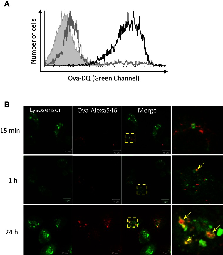
Figure 3.
Adherent HK mononuclear phagocytes hydrolyze Ova and accumulate the Ag within acidified endocytic compartments. (A) Head kidney leukocytes were incubated for 4 h with 5 μg/ml of Ova-DQ. Non-adherent and adherent cells were harvested and analyzed separately with flow cytometry. Gray contour – non-adherent MHCII++ granulocytes, black contour – adherent MHCII+ cells. The filled contour shows a non-stained control. Ova-DQ fluorescence is detected in the green channel. Similar results were obtained with cells from two individuals. (B) Adherent head kidney mononuclear phagocytes attached to chambered coverglass slides were stained with LysoSensor Green (pKa 5.2) and incubated with Ova-Alexa647 for the indicated periods prior to live imaging using a confocal microscope. The arrows in the magnified overlap regions indicate accumulation of Ova within late endosome/lysosome compartments.

