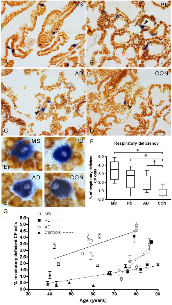
Figure 1. Respiratory deficient cells in the choroid plexus.
A–E: Sequential complex IV (cytochrome c oxidase or COX) and complex II (succinate dehydrogenase or SDH) histochemical assays identified cells with intact complex IV activity [respiratory efficient cells, stained brown] as well as lacking complex IV but with intact complex II [respiratory deficient, stained blue] within choroid plexus (CP) from multiple sclerosis (MS, a), Parkinson’s disease (PD, b), Alzheimer’s disease (AD, c) and control (CON, d) cases. Respiratory deficient cells (blue) were located in the CP epithelium and were larger than respiratory efficient cells (brown) in all four groups (ei-iv), which is consistent with reports of oncocytic cells in CP. Scale bar; 50µm in a–d and 10µm in e. Cases shown are MS6 (age 59), PD1 (age 76), AD1 (age 72) and CON5 (age 60).
F: The proportion of RD cells as a percentage of all CP epithelial cells was significantly greater in MS (3.39% ± 1.08, *p<0.001), PD (2.45% ± 1.20, #p=0.007) and AD (1.91% ± 0.99, †p=0.02) compared with controls (0.82% ± 0.52, Kruskal Wallis test p<0.001). MS choroid plexus contained significantly greater proportion of respiratory deficient epithelial cells than AD (p=0.027), despite significantly lower mean age of MS cases compared with AD (see Table 1). The difference in the density of respiratory deficient cells between MS and PD CP was not statistically significant. A mean of 1327±146 cells, with a nucleus stained by Hoechst, were included per case.
G: When the density of respiratory deficient cells in the choroid plexus was plotted against age, we observed a significant correlation between the two parameters in MS (p=0.037 and r2=0.439), AD (p=0.049 and r2=0.775) and controls (p=0.037 and r2=0.485). There was a trend towards an increase in the density of respiratory deficient cells with age, which was not statistically significant, in PD, where the age range was narrow (Table 1).

