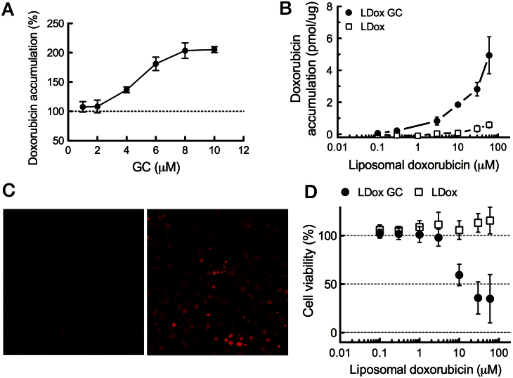
Figure 4. GC enhances cellular accumulation of doxorubicin in mouse mammary tumour (WEP) cells and restores cytotoxicity.
(A–C) Intracellular doxorubicin accumulation in WEP cells in presence or absence of GC. WEP3 cell cultures were derived from Ecad−/−;p53−/− mammary tumours and were pre-incubated with free GC from an ethanol solution, followed by 1 hour free doxorubicin incubation (50 μM) (A) Intracellular doxorubicin accumulation was quantified by fluorometry on cell lysates and normalized against control (no GC pre-incubation). (B) Doxorubicin accumulation in WEP3 cells incubated for 24 hours with liposomal doxorubicin plus GC (LDox GC), or conventional doxorubicin liposomes (LDox) as control. (C) Fluorescence micrographs of doxorubicin, taken at equal exposure times, on (live) WEP3 cells immediate after 24 hours incubation with LDox GC (right panel) or LDox (left panel) (10 μM). (D) WEP3 cell viability after 24 hour incubation with LDox GC or LDox. Cells were kept in culture for an additional 48 hours. Viability was then assessed by the metabolic XTT assay. Data are expressed as mean percentages to untreated cells (SD, n = 6). As a control, incubation with the same liposomes but devoid of doxorubicin did not affect cell viability.

