Figure 2.
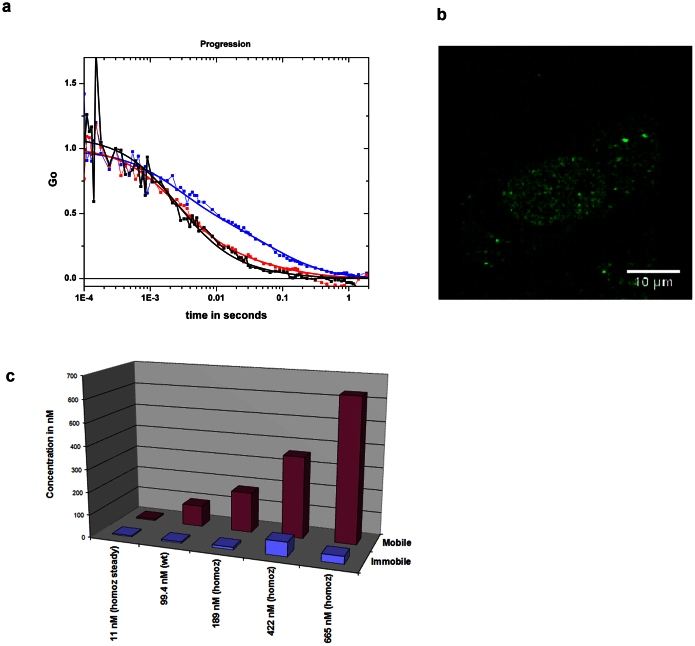
Increased MYC augments mobile-MYC (a) ACF of three MEF cells with different concentrations of eGFP-cMyc. The concentrations are 10 (blue), 94 (red) and 400 nM (black). All three ACFs have been normalized to G(blue) at τ = 1e − 3. (b) Image of MEF cell at 970 excitation containing about ~50 nM of eGFP-cMyc (high resolution). (c) The fractions of the mobile and immobile populations obtained from transfections of wild type and homozygous cells are shown.

