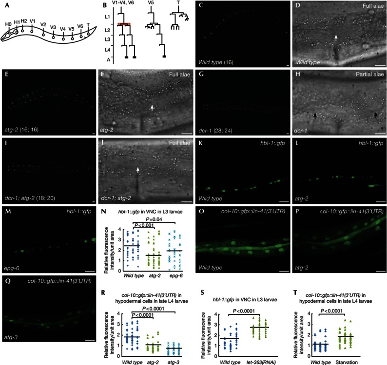
Figure 1.
Loss of autophagy activity suppresses the retarded heterochronic defects in miRISC mutants. (A) Schematic illustration of seam cells in a wild-type L1 larva. (B) Division pattern of seam cells at the postembryonic stage in hermaphrodites. Seam cells V1–V4 and V6 divide symmetrically at the L2 stage (highlighted in red), thus doubling in number. (C–F) At the young adult stage, wild-type and atg-2 mutant animals have 16 seam cells on each side, visualized by the seam cell-specific marker scm::gfp. Cuticular alae (arrow) run continuously from anterior to posterior. The number of seam cells is shown in brackets. (G,H) dcr-1 young adults have an increased number of seam cells (shown in brackets) and gaps in the alae (between arrows). (I,J) atg-2 mutations suppress the retarded heterochronic defect in dcr-1 mutants. Complete alae (arrow) are formed in dcr-1; atg-2 double mutants. (K–M) Confocal images showing expression of hbl-1::gfp::hbl-1 reporter in VNC in wild type (K), atg-2 (L) and epg-6 (M) L3 larvae. (N) Distribution of relative fluorescence intensity of HBL-1::GFP in 50 unit areas in VNC in the indicated strains. (O–Q) Confocal images showing the expression of the col-10::gfp::lin-41(3′UTR) reporter in wild type (O), atg-2 (P) and atg-3 (Q) late L4 larvae. (R) Distribution of relative fluorescence intensity of col-10::gfp::lin-41(3′UTR) in 50 unit areas of hypodermal cells in the indicated strains. (S,T) Distribution of relative fluorescence intensity of hbl-1::gfp in VNC and col-10::gfp::lin-41(3′UTR) in hypodermal cells in 50 unit areas in wild type, let-363(RNAi) or starvation-treated animals. Black lines in (N,R–T) represent the average fluorescence intensity of the reporter in each strain. P values are determined by Student’s two-tailed unpaired t-test in (N,R–T). (C,E,G,I) Scale bar, 20 μm; (D,F,H,J–M,O–Q) scale bar, 10 μm. GFP, green fluorescent protein; miRISC, miRNA-induced silencing complex; RNAi, RNA-mediated interference; UTR, untranslated region; VNC, ventral nerve cord.

