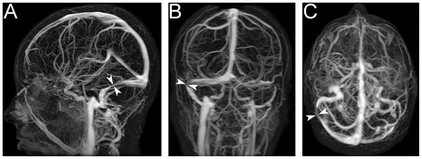
Figure 4. Transverse cerebral venous sinus stenosis in an IIH patient, as seen on contrast-enhanced MR venography. There is a long narrow stenosis of the right transverse venous sinus (arrowheads) and a hypoplastic left transverse venous sinus.

A: Sagittal plane.
B: Coronal plane.
C: Axial plane.
