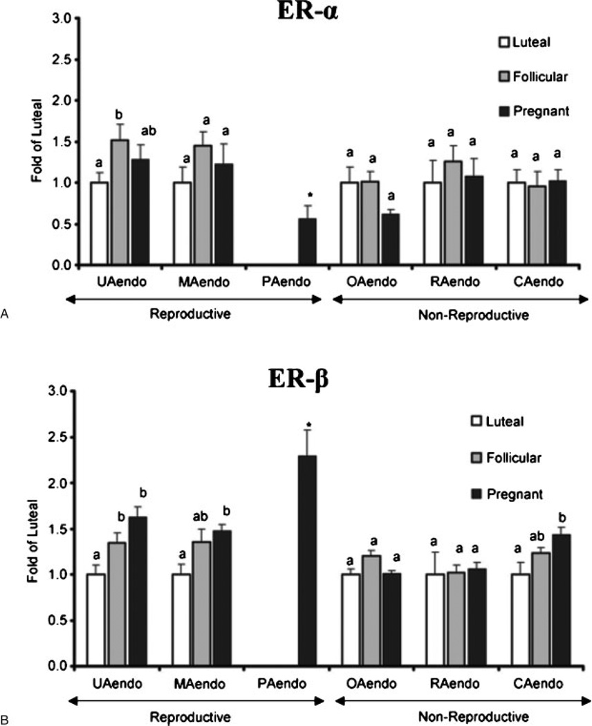
Figure 3.
(A) Estrogen receptor (ER)-α and (B) ER-β protein expression in reproductive versus nonreproductive arterial endothelia. Western blot analysis was performed to evaluate the relative levels of ER-α and ER-β protein in luteal, follicular, and pregnant sheep reproductive and nonreproductive endothelia. Samples from luteal, follicular, and pregnant sheep are expressed as fold of the average of all luteal samples within an artery type, run on the same Western blots. Expression of PAendo is given as fold of the average luteal UAendo was also run on the same blot. Treatment groups: luteal: uterine (n = 12), mammary (n = 5), omental (n = 7), renal (n = 6), and coronary (n = 7). Follicular: uterine (n = 8), mammary (n = 5), omental (n = 8), renal (n = 6), and coronary (n = 8). Pregnant: uterine (n = 12), mammary (n = 6), placental (n = 8) omental (n = 8), renal (n = 8), and coronary (n = 8). Data are means plus or minus standard error of the mean. Means with different letters are statistically different (p < 0.05) within a tissue preparation. For ER-α UAendo: Lut < Fol (p < 0.05), For ER-β UAendo: Lut < Fol (p < 0.05) and Lut < Preg (p < 0.001); MAendo: Lut < Preg (p < 0.01); CAendo: Lut < Preg (p < 0.05). *For ER-α: PAendo < Luteal UAendo but for ER-β PAendo > Luteal UAendo (p < 0.05).35 (With permission from Byers MJ, et al. J Physiol 2005;565:85–99.)

