Figure 2.
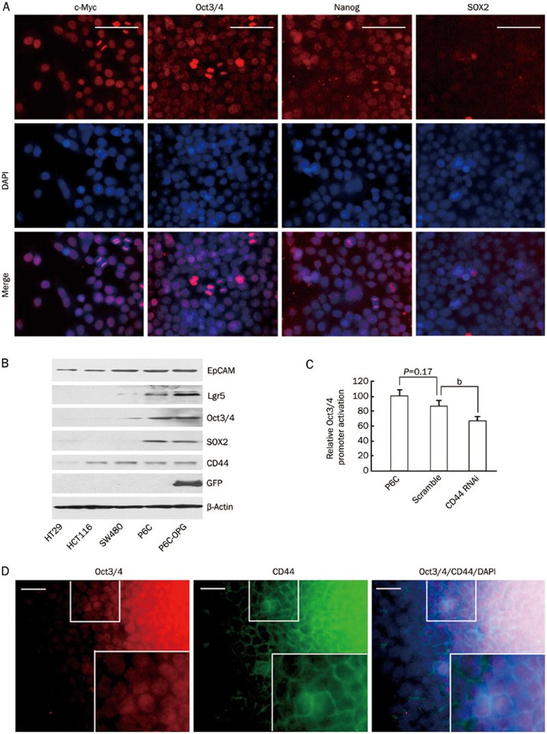
Stemness genes expression in P6C cells. (A) Immunofluorescence of stemness proteins in cultured P6C cells. P6C spheres were dissociated, seeded onto coverslips and allowed to attach for 3 h. After fixing cells with paraformaldehyde, the cells were incubated with the indicated antibodies. DAPI was used for nuclei counter staining. Scale bars, 100 μm. (B) Stemness gene expression detected by Western blotting. P6C cells were stably transfected with the pOct3/4 promoter-EGFP (OPG) construct. The whole cell lysates of HT29, HCT116, SW480, P6C, and P6C-OPG cells were loading equally, subjected to SDS-PAGE and transferred to nitrocellulose membranes. The membranes were then incubated with the indicated antibodies and visualized using an ECL system. (C) Effect of CD44 shRNA on Oct3/4 expression in P6C cells. Relative Oct3/4 promoter activity was measured by GFP fluorescence intensity as detected using flow cytometry. P6C and parental cells were used as controls. Each sample was performed in triplicate, and the experiment was repeated 3 times. bP<0.05. (D) Co-expression of endogenous Oct3/4 and CD44 in a P6C single-cell derived clone. Magnified images are shown in the lower right panel. Scale bars, 50 μm.

