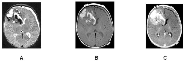
Figure 3.

Hemorrhagic primitive neuroectodermal tumor of the right frontal lobe in a 4-month-old infant. Axial T2 (A), axial T1 pre-contrast (B), and post-contrast (C) images show a large mass with significant hemorrhagic products (bright T1 signal before contrast and mixed low and bright T2 signal). The nonhemorrhagic more peripheral components enhance mildly (C). There is no surrounding edema. A subdural hematoma is evident on the left.
