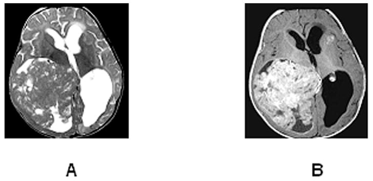
Figure 5.

Axial T2 (A) and contrast-enhanced T1 (B) images of an 8-month-old with a large rhabdoid tumor arising in the posterior right lateral ventricle. The mass shows intense enhancement, and has low T2 signal with small islands of high T2 signal that do not enhance (small cysts or necrotic foci). A metastatic lesion is evident deep to the left anterior insula. Hydrocephalus results from posterior 3rd ventricular compression by the tumor.
