Figure 1.

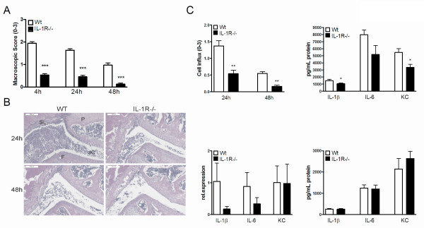
Murine Lyme arthritis is IL-1-dependent. (A) Macroscopic score of the knees in either wild-type (WT) (white bars), or IL-1 Receptor-/- mice (black bars). After 4, 24, and 48 hours of intra-articular (i.a.) injection of 1 × 10^7 live B. burgdorferi, at least 10 knees per group. Data are mean ± SEM from eight animals in each group; ***P <0.0001; Mann-Whitney U test, two-tailed. (B) Murine Lyme arthritis in WT, or IL-1R-/- mice. Histology (H&E staining) 24, and 48 hours after i.a. injection of B. burgdorferi in knee joints. 200× magnification; P, patella; F, femur; JC, joint cavity; SL, synovial lining. Scale bar represents 100 μM. (C) Upper left: Scored cell influx after 24 and 48 hours of i.a. injection with B. burgdorferi. Upper right and lower left: 4 hours after i.a. injection of 1 × 10^7 live B. burgdorferi in 10 μL of PBS, patellae were cultured for 1 h and IL-1β, IL-6 and keratinocyte-derived chemokine (KC) protein levels and mRNA expression levels were measured using Luminex and qPCR, respectively. Lower right: After 24 hours of infection, 1 × 10^5 peritoneal macrophages were stimulated for 24 hours with live B. burgdorferi. White bars represent cytokine induction by WT mice, black bars are IL-1R gene-deficient mice, at least five animals/group. *P <0.05, **P <0.01; Mann-Whitney U test, two-sided.
