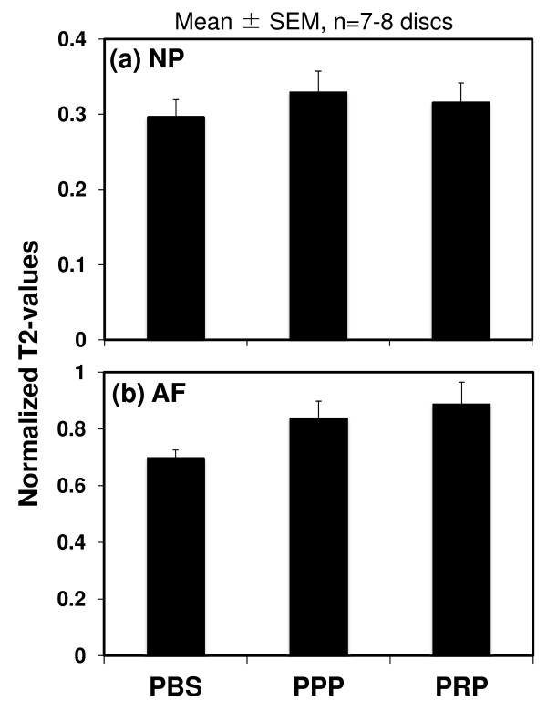
Figure 4.
Magnetic resonance imaging results of normalized T2 values. Magnetic resonance imaging (MRI) analysis was performed at 8 weeks after the phosphate-buffered saline (PBS) (control), platelet-poor plasma (PPP)-releasate, and platelet-rich plasma (PRP)-releasate injections. The normalized T2 value of the PBS group was the lowest on average, but no statistical significance was found. Values are shown as mean ± standard error of the mean (SEM): PBS; n = 7 discs, PPP; n = 7 discs, PRP; n = 8 discs.

