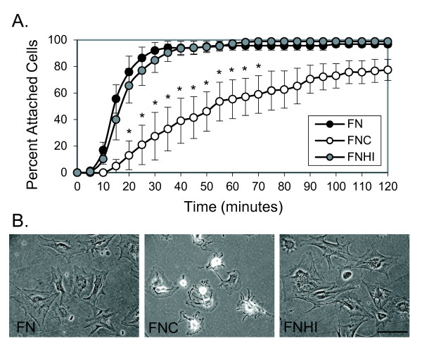
Figure 2.
Murine synovial fibroblasts have impaired attachment and spreading on citrullinated fibronectin. (A) Murine synovial fibroblasts were plated on dishes coated with fibronectin (FN), citrullinated fibronectin (FNC), or fibronectin incubated with heat-inactivated peptidyl arginine deiminase (FNHI) and were imaged using time-lapse microscopy at 10× magnification. The percentage of cells attached in a 10× field was determined every 5 minutes. Graph depicts average and standard error at each time point (n = 4 experiments). *P < 0.05. (B) Representative images from (A) taken at 2 hours at 20× magnification. Bar = 100 μm.

