
Figure 1.
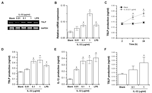
IL-32-induced mRNA expressions and protein production of TSLP in THP-1 cells. THP-1 cells (3 × 10⁶) were treated with IL-32 (0.01, 0.1, and 1 μg/ml) or LPS (10 ng/ml) for 5 h. The TSLP mRNA expression was measured by the RT-PCR (A) and real-time PCR (B) method. (C) THP-1 cells (1 × 10⁵) were stimulated with IL-32 (0.1 μg/ml) for various times. (D) THP-1 cells (3 × 10⁵) were stimulated with IL-32 (0.01, 0.1, and 1 μg/ml) or LPS for 24 h. The production of TSLP in the supernatant was measured by the ELISA method. (E) The production of IL-1β in the supernatant was measured by the ELISA method. (F) PBMCs (3 × 10⁵) were stimulated with IL-32 (0.1, and 1 μg/ml) for 24 h. The production of TSLP in the supernatant was measured by the ELISA method. Each datum represents the mean ± SEM of three independent experiments. Blank, unstimulated cells. *P < 0.05; significantly different from unstimulated cells' value. ELISA, enzyme-linked immunosorbent assay; IL, interleukin; LPS, lipopolysaccharide; PBMC, peripheral blood mononuclear cells; RT-PCR, reverse transcriptase polymerase chain reaction; TSLP, thymic stromal lymphopoietin.