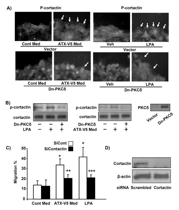
Figure 5. PKCδ regulates ATX-V5-induced cellular migration.
A) A549 cells were scratched, and then were treated with ATX-V5 Med or LPA (1 μM) for 3 h. Immunostaining of phosphorylated PKCδ was performed using an antibody to p-PKCδ. Arrows indicate phospho-PKCδ. Also shown are representative images from three independent experiments. B) Cells were infected with Dn-PKCδ adenovirus or Dn-PKCζ or Dn-PKCα adenovirus (10 MOI) for 24 h, then cells were incubated with ATX-V5 Med and cell migration was measured by the scratch assay. The data represents mean ± S.D. from three independent experiments. *p<0.05, compared to Cont Med; **p<0.05, compared to ATX-V5 Med alone.

