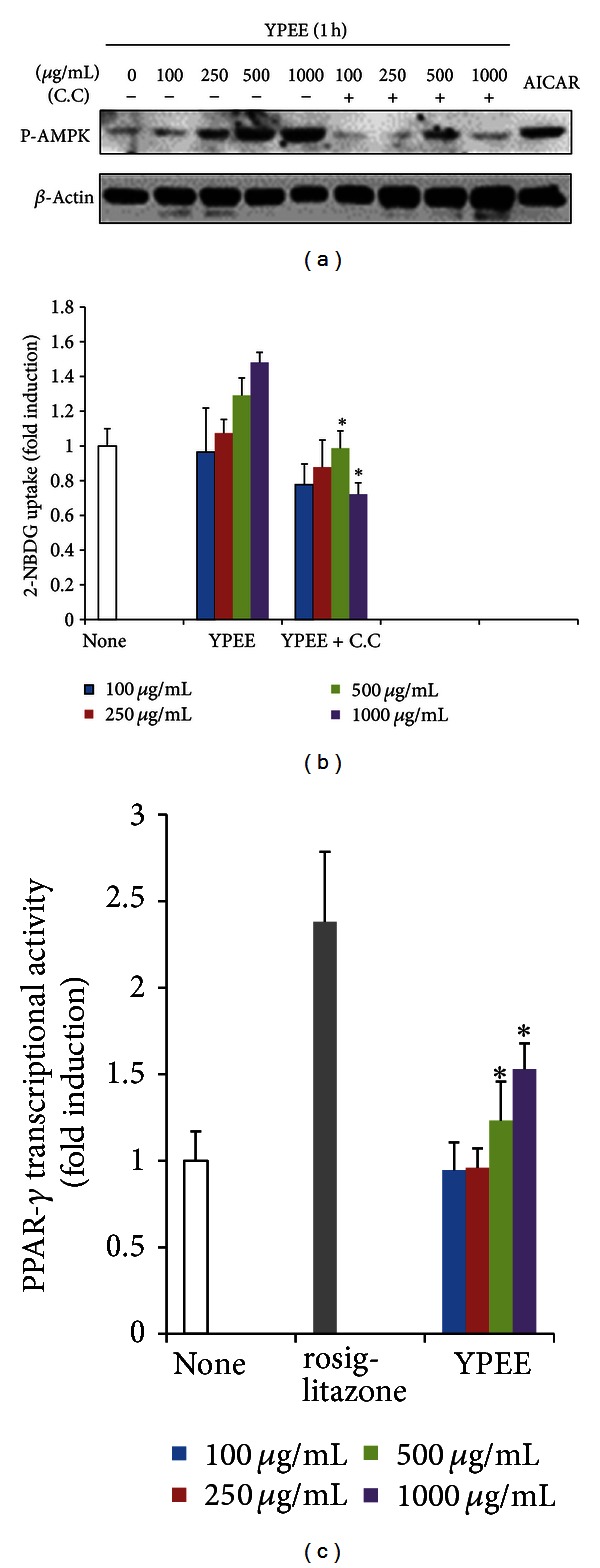
Figure 2.

Effect of YPEE on AMPK and PPAR-γ. Cells were pretreated with 10 μM C.C for 30 min and consecutively exposed to YPEE for 1 h in a dose-dependent manner. Western blot analysis was performed with phosphospecific AMPK and normal beta-actin antibodies (a). Cells were pretreated with 10 μM C.C for 30 min and consecutively exposed to YPEE, and then, the 2-NBDG uptake assay was performed, as described in Section 2 (b). Data are expressed as mean ± SD. *P < 0.05 versus YPEE. PPAR-γ expression vector and PPRE-luc vectors were cotransfected in HEK293 cells and exposed to YPEE or 25 μM rosiglitazone. PPAR-γ transcriptional activity was measured with the luciferase assay system (c). Data are expressed as mean ± SD. *P < 0.05 versus none.
