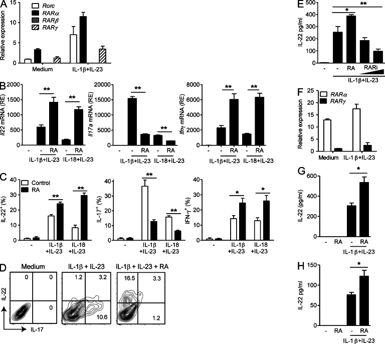
Figure 1.
RA enhances IL-22 production by γδ T cells and ILC3. (A) Relative mRNA expression of Rorc, Rarα, -β, and -γ in purified γδ T cells from LNs, ±IL-1β and IL-23 stimulation for 48 h. (B) Relative mRNA expression (RE) of Il22, Il17a, and Ifnγ in LN γδ T cells stimulated with IL-1β and IL-23 or IL-18 and IL-23 with or without RA. (C) IL-22, IL-17, and IFN-γ production by ICS on purified LN γδ T cells stimulated with IL-1β and IL-23 or IL-18 and IL-23 ± RA or vehicle control for 72 h (mean ± SE). (D) ICS on purified LN γδ T cells stimulated with IL-1β and IL-23 ± RA. (E) IL-22 production by ELISA on purified LN γδ T cells stimulated with IL-1β and IL-23 for 72 h ± 100 nM RA or 0.5 or 5.0 µM RARi (mean ± SD). (F) Relative mRNA expression of Rarα and -γ in FACS-sorted lamina propria NCR+ ILC3 (CD3−CD19−CD11c−NK1.1−NKp46+) with and without stimulation with IL-1β and IL-23 for 48 h. (A, B, and F) Results are mean and SD values for triplicate samples. (G and H) IL-22 production detected by ELISA on lamina propria NCR+ ILC3 (G) or γδ T cells (H) stimulated with IL-1β and IL-23 ± RA (mean ± SD). Results are representative of two to four independent experiments (n = 3 for A, B, E, and F; n = 4 for C, G, and H; D is representative of four samples). *, P < 0.05; and **, P < 0.01 versus DMSO control.

