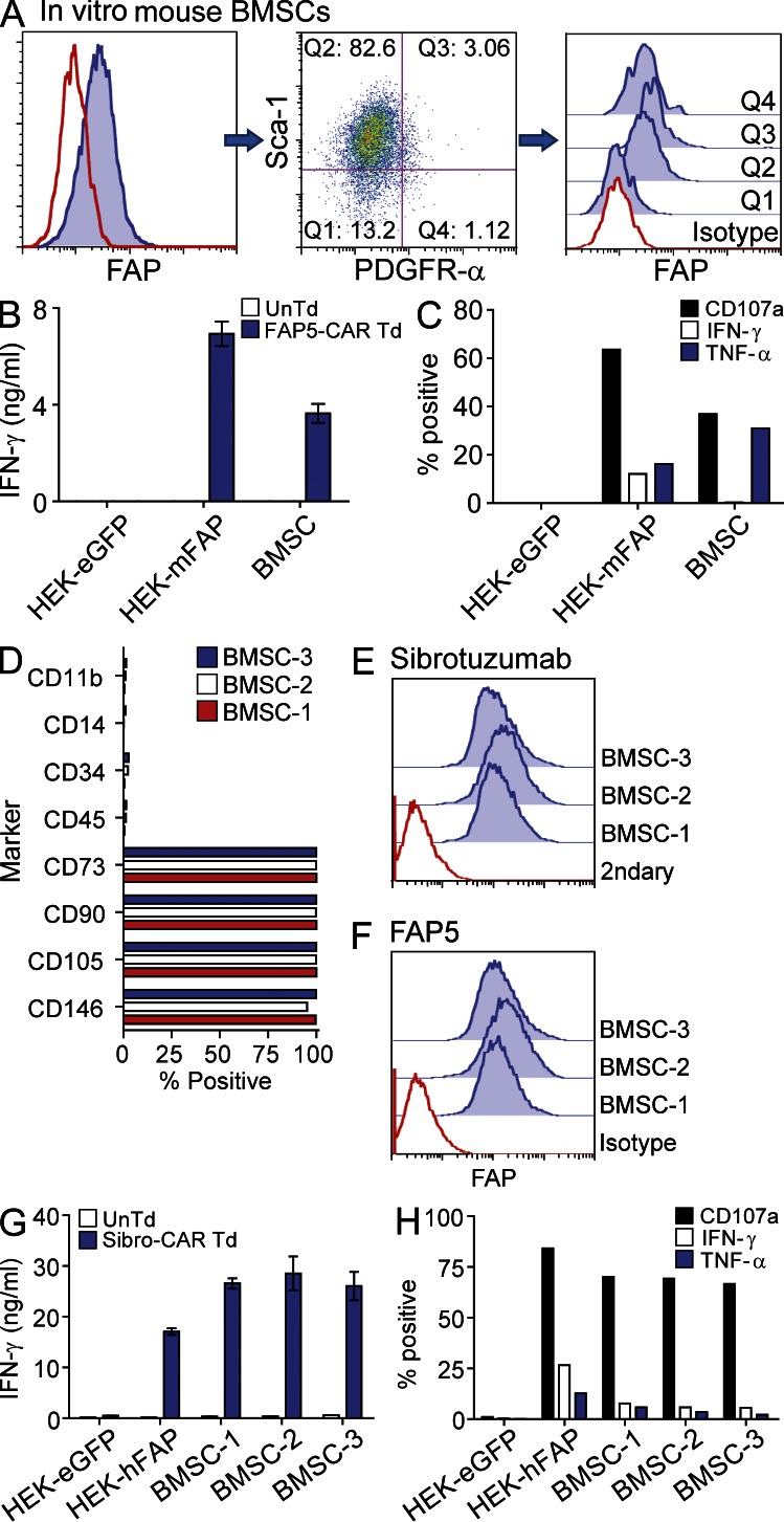
Figure 5.
Murine and human multipotent BMSCs express FAP and are recognized by T cells expressing FAP-reactive CARs. Passage-5 in vitro–expanded murine BMSCs were stained with antibodies specific for Sca-1, PDGFR-α, and FAP, and assessed by flow cytometry (A). “Q” represents quadrant. Solid lines are isotype controls and filled histograms are FAP stained. UnTd or FAP5-CAR Td T cells were cultured overnight with murine BMSCs and supernatants were assessed for IFN-γ by ELISA (B), and cells were further analyzed for expression of CD107a and production of IFN-γ and TNF by ICS (C). Mean ± SD. Data are representative of two independent experiments. Flow cytometric phenotype of in vitro–expanded human BMSCs derived from three different donors (D). BMSCs from D were stained with the FAP-specific monoclonal antibodies Sibrotuzumab (E) and FAP5 (F) and assessed by flow cytometry. Solid lines are isotype or secondary antibody controls and filled histograms are FAP or Sibrotuzumab stained. UnTd or Sibro-CAR Td T cells were cultured overnight with BMSCs and the supernatants assessed for IFN-γ by ELISA (G), and cells were further analyzed for CD107a expression and IFN-γ and TNF production by ICS (H). Mean ± SD. Similar results were seen with two additional T cell donors.

