
Figure 5.
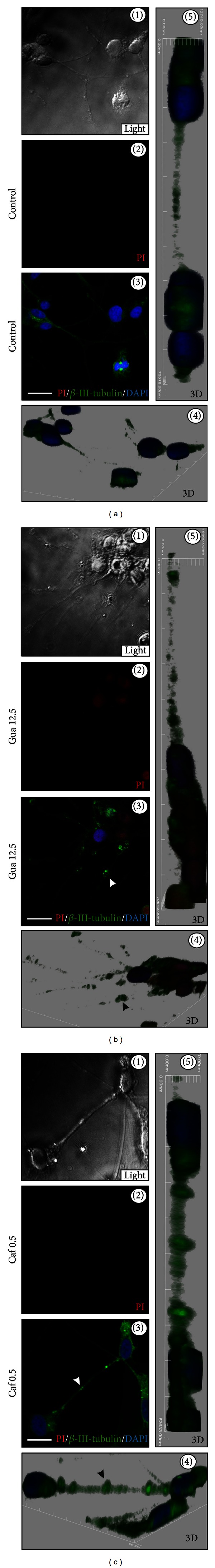
Qualitative analysis of drug-induced neuritic morphology. Confocal immunofluorescence microscopy (CM) of β-III-tubulin expression (green fluorescence), propidium iodide (PI) (red fluorescence), and DAPI (nuclei) on human neuronal SH-SY5Y cells (a), after treatment with 12.5 mg/mL of guarana (b) and 0.5 mg/mL of caffeine (c) for 4 hours in FBS-free culture medium. White and black arrow heads (in 2D and 3D representations) mark the presence of neuritic swelling after guarana (12.5 mg/mL) and caffeine (0.5 mg/mL) treatments. Scale bars represent 10 μm.