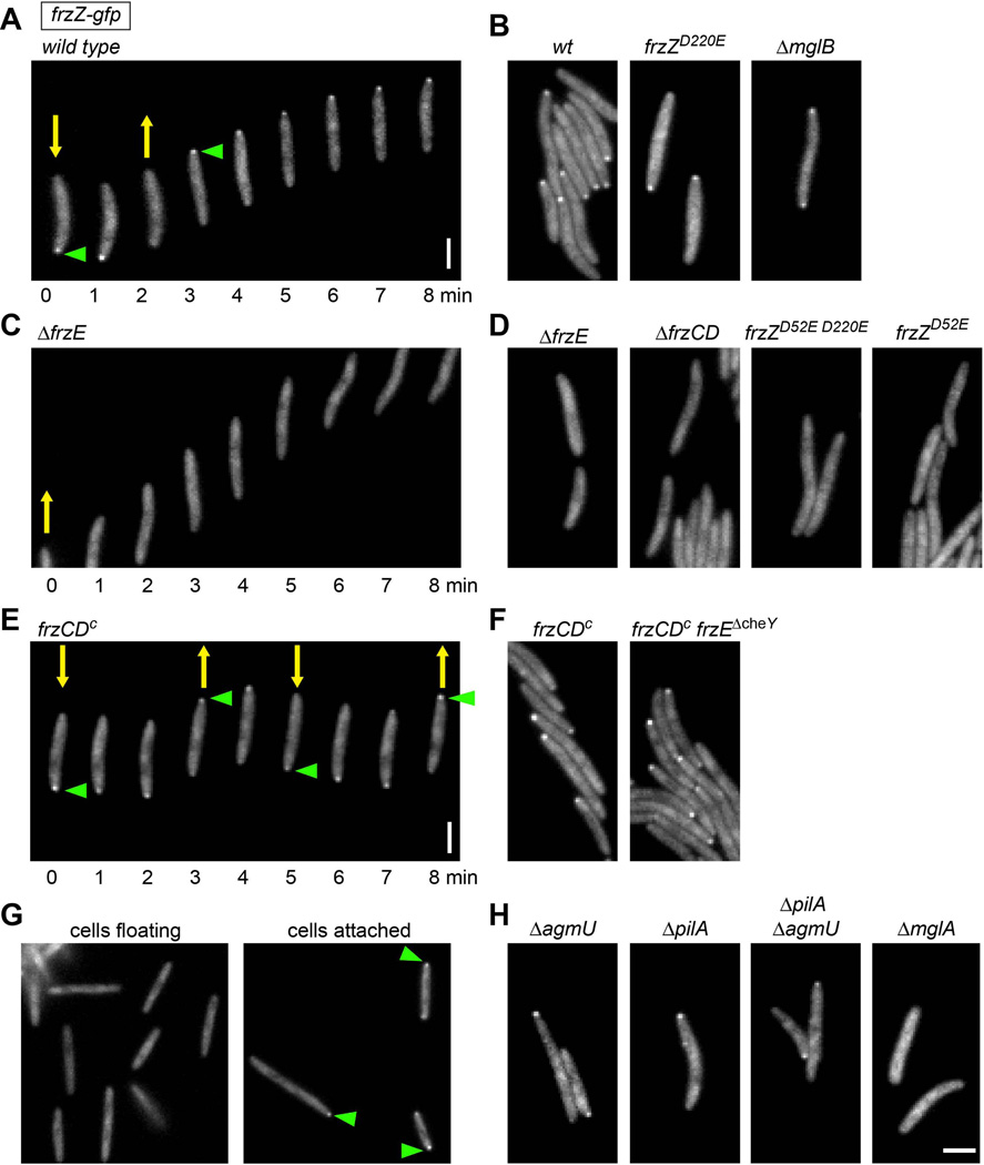
Figure 6.
Fluorescence microscopy of FrzZ-GFP in live M. xanthus cells reveals distinct and phosphorylation-dependent localization at the leading cell pole. Yellow arrows indicate the direction of cell movement. Green arrowheads indicate FrzZ-GFP accumulation at the leading cell pole. A, B. strains that show wild type levels of FrzZ phosphorylation. C, D. strains that shown no FrzZ phosphorylation. E, F. strains with elevated FrzZ phosphorylation levels. G. hyper-reversing strain frzZ-gfp frzCDc was grown in liquid culture fro 72 h and imaged after transferring cells to a glass slide. Left panel: floating cells; right panel: cells attached to surface. H. strains affected in social or gliding motility. Images were taken every 1 min for time-lapse acquisition (A, C, E). Scale bars represent 2 µm.

