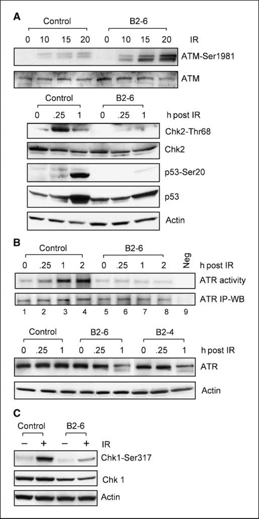
Figure 5.
The effect of BRCA1 expression on IR-induced ATM and ATR signalings. A, top, MCF-7 cells expressing Luc-shRNA (Control) or BRCA1-shRNA (B2-6) were treated with 10-Gy IR, incubated for 15 min, and assessed for ATM-Ser1981 phosphorylation and total ATM by immunoblotting. Bottom, cells were treated as described above, incubated for the indicated times, and then analyzed for phosphorylation of Chk2-Thr68 and p53-Ser20 by immunoblotting. ATM, Chk2, p53, and actin levels in the lysate samples were determined by immunoblotting. B, top, Cells were exposed to 10-Gy IR and incubated for the indicated times. ATR kinase was immunoprecipitated from 500-µg cell lysate and analyzed for ATR activity using recombinant p53 protein as substrate (ATR activity, lanes 1–8), as described in Materials and Methods. As a negative control, ATR activity in irradiated cells was assayed using GST recombinant protein as substrate (ATR activity, lane 9). ATR levels in all immunoprecipitates were assessed by immunoblotting using anti-ATR antibody N-19 (ATR IP-WB). Bottom control (Control) and BRCA1-shRNA–expressing cells (B2-6 and B2-4) were exposed to 10-Gy IR, incubated for the indicated times, and analyzed for ATR protein levels by immunoblotting (ATR). C, cells treated with or without 10-Gy IR were incubated for 1 h and then analyzed for levels of Chk1-Ser317 phosphorylation and total Chk1.

