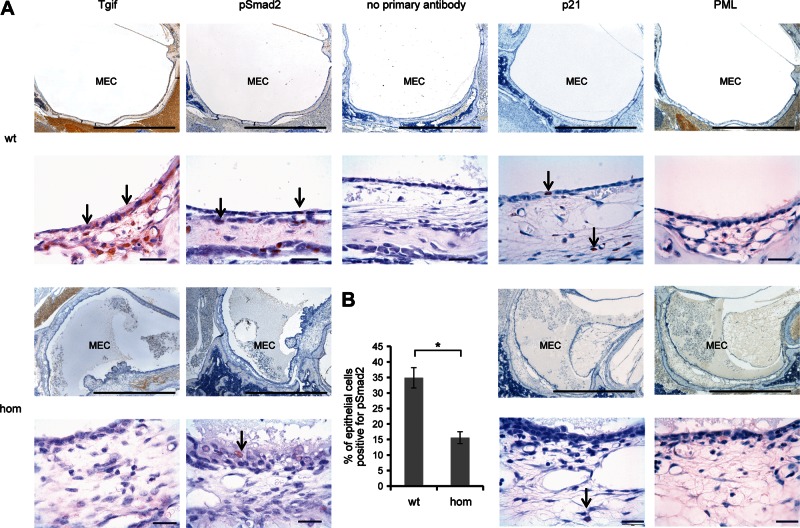
Figure 9.
Protein expression in middle ear epithelia in Tgif mutant mice. (A) Immunohistochemistry of middle ear sections of wild-type (wt, +/+) and homozygous (hom, Tgif/Tgif) mice, age 21 days after birth, stained with TGIF, phosphoSmad2 and p21 and cPML antibodies. Arrows indicate nuclear localization in the epithelial cells. cPML was localized in the cytoplasm of the cells. No difference in the localization between wild-type and homozygous cells was observed with the cPML antibody. MEC, middle ear cavity. Scale bars = 1 mm and 20 μm. (B) Graphic comparison of the percentage of epithelial cells in the middle ear positive for pSmad2. Bars: standard error of mean. *P < 0.05.

