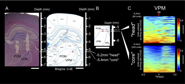
Figure 5.
Both head and core of barreloids in VPM present anticipatory neural activity. A, Histological verification (left) and comparison with standard diagrams (right) (Paxinos and Watson, 1998) of microelectrode placement in the POM and VPM. The white lines in the rulers and the two red markings indicate the depths at which neural activity corresponding to the head (−5.2 mm) and core (−5.4 mm) regions of the VPM was recorded. B, Magnification of standard diagram showing the depths used to define head and core of the barreloids. C, Top and bottom show the neural ensemble activity recorded from depths corresponding to head and core of the barreloids. Each row in each panel represents the activity of a single unit during a session normalized to its maximum firing rate. Each of the different colors represents a significant variation in the firing rate, with red indicating excitation, and deep blue indicating inhibition. Time 0 corresponds to the discrimination bars BB. Units were ordered by the maximum firing rate in −0.5 to 0 s. In both the head and core of the VPM, the bottom rows of neurons exhibited increased anticipatory firing rates immediately before the whiskers contacted with the discriminanda (time 0). This pattern of anticipatory increased activity was more pronounced in the head of the barreloids than in the core. In the core of the VPM, the period of anticipatory activity was mainly characterized by a strong inhibition before and after the whiskers sampled the aperture bars. During the discrimination period, a marked increase in firing activity was present in the core of the VPM. A similar increase was not as evident in the head of the VPM. These results show that anticipatory firing could be found at all depths studied in the VPM, but that each of the two different compartments of this thalamic nucleus displayed a very specific pattern of firing modulation. In the head of the VPM, the pattern of activation was closer to the one described for M1, POM, and S1 infragranular layers, while in the core of the VPM, the pattern was closer to the one observed in granular layer of S1. VPL, Ventral posterior lateral thalamic nucleus.

