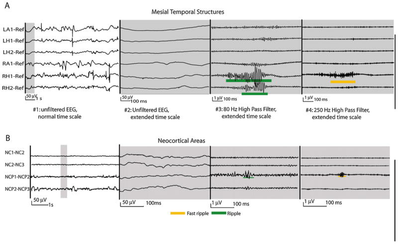
Fig. 2.
HFO recorded with macroelectrodes. This figure demonstrates ripples and fast ripples in the mesial temporal (A) and neocortical structures (B). HFOs are visualized on a different time scale and with different filter settings than the usual clinical EEG. In the two EEG segments on the left the amplitude scale is 50 times increase compared to those on the left to demonstrate the very small scale HFOs. HFOs in the mesial temporal structures are larger, of higher amplitude and more frequent than those in neocortical areas. Both EEGs derived from patients with non-lesional epilepsies.
Source: Figure adapted from Jacobs et al. (2009c) with permission from John Wiley & Sons.

