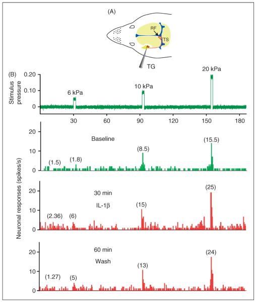
Figure 1.
Experimental arrangement and raw data example of the responses of one C-unit meningeal nociceptor to local application of IL-1β. (A) Schematic localization of the recording site for the meningeal nociceptors in the trigeminal ganglion (TG) and the mechanical receptive field (RF) of the recorded neuron on the left transverse sinus (TS). (B) Peri-stimulus time histograms (0.5 s beans) depicting the responses to threshold and suprathreshold mechanical stimulation of the unit's dural receptive field as well as ongoing discharge rates at baseline (green trace), and during the sensitization stage following 30 min of IL-1β administration and 60 min after wash with SIF (red traces). The numbers in parenthesis indicate mean spikes/s. Note the persistent sensitization to mechanical stimulation despite 60 min of wash. IL-1β: interleukin 1β, IL-6: interleukin 6, SIF: synthetic interstitial fluid.

