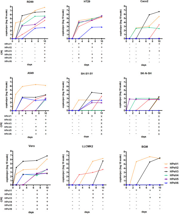
Figure 1.
Replication kinetics of HPeV1 to 6. Growth kinetics of laboratory adapted HPeV1 to 6 on different cell lines including appearance of CPE at days 0, 1, 3, 7 and 10. Cells were infected with HPeVs at a MOI 0.001 and viral RNA was detected in the supernatant with RT-PCR at days 0, 1, 3, 7 and 10. The 10log virus copies were calculated with a standard curve, and the input virus copies per PCR at day 0 was subtracted. HPeV1: orange line, HPeV2: red line, HPeV3: black line, HPeV4: green line, HPeV5: yellow line, HPeV6: blue line. CPE was scored as positive (+), negative (−) and dead cells (X).

