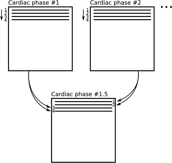
Figure 10.

View-sharing. An intermediate cardiac-phase (bottom) is constructed from the acquired data of the first and second cardiac-phases (top). In this diagram only one k-space segment with 4 phase-encode lines is shown, this process is repeated for all subsequent segments.
