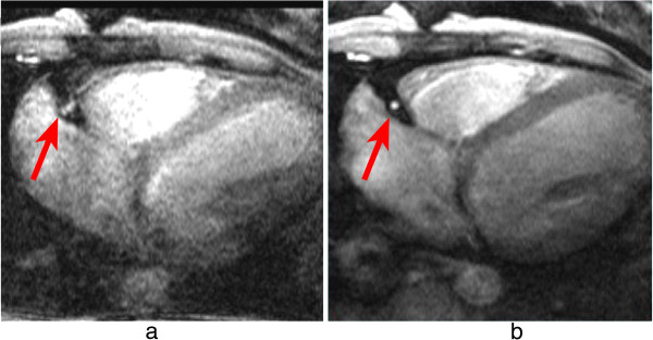
Figure 18.

Right coronary imaging with cardiac motion artefacts. Magnitude images from a phase-velocity mapping study of the right coronary artery (arrows) acquired during systole. a) Breath-hold with long acquisition window during each cardiac-cycle, b) Retrospective respiratory gated with shorter acquisition window during each cardiac-cycle. Image a is considerably degraded due to cardiac motion in the longer acquisition window. (Adapted and reprinted, with permission, from reference [50]).
