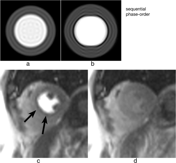
Figure 19.
Myocardial perfusion and cardiac motion artefacts (sequential phase-order). a) Numerical simulations of a short-axis image (including Gibbs ringing) with no in-plane motion. b) Same as a but with in-plane cardiac motion (myocardial radial contraction) with a sequential phase-order acquisition. c) In vivo short-axis image of a perfusion scan with a bSSFP sequence with a sequential phase-order acquisition. A subendocardial dark rim artefact is visible likely to be a superposition of motion and Gibbs ringing artefacts (arrows). d) Same as c but after first-pass; the contrast between the LV and myocardial signal is reduced and the dark rim artefact is no longer visible.

