


Figure 22.
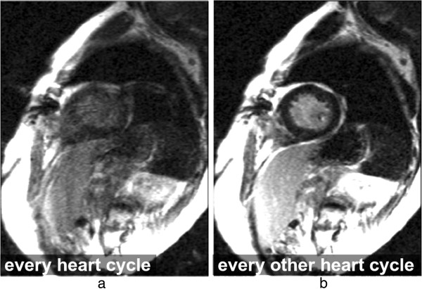
Arrhythmia artefacts in LGE. LGE short-axis image, in a patient with arrhythmia. a) Image acquisition for every heart-cycle. b) Image acquisition for every other heart-cycle. Image quality is improved in b by acquiring data at every other heart-cycle only, reducing the contrast inconsistencies between different k-space segments that were created by the irregular heart-rate.