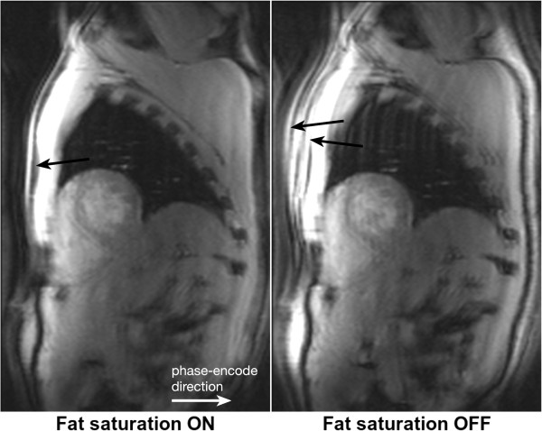
Figure 31.
h-EPI chemical shift artefact. Short-axis images acquired with a centric interleaved h-EPI sequence with fat-saturation preparation turned on and off. When fat saturation pulses are used, the fat signal in the chest wall is efficiently suppressed (arrow). When there is no fat-saturation preparation, the fat signal in the chest wall is visible and, due to chemical shift, is displaced approximately 4 pixels in both directions (arrows).

