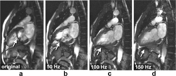
Figure 39.

bSSFP and B0 inhomogeneities: tuning frequency. a-d) Series of vertical long axis bSSFP images acquired with different tuning frequency offsets: original (0Hz), 50Hz, 100Hz, and 150Hz. Black band and flow artefacts through the ventricle are shown (arrows). As the frequency is adjusted the artefacts are shifted away from the heart. Image d shows another dark band approaching the heart from the top (dotted arrow), as the tuning frequency is shifted. This approaching frequency-offset causes the blood signal to be hyperintense in the image plane, similar to that shown in Figure 38b) (227 ms).
