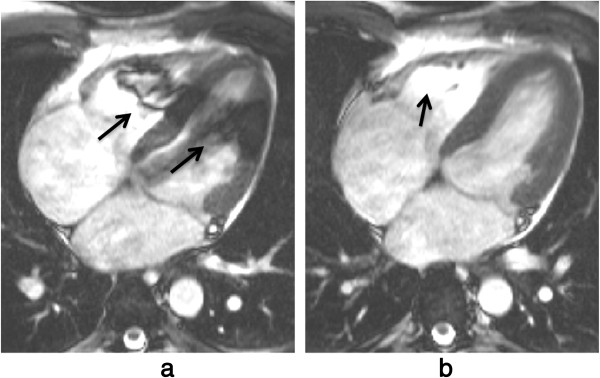
Figure 40.

bSSFP and B0 inhomogeneities: tuning frequency II. a) Long-axis bSSFP cine frame showing dark bands in the heart (arrows) due to field distortions caused by an air pocket in the colon (not shown in this plane). b) The same frame after a manual frequency tuning. The shift of the scanner centre-frequency removed the artefact from the LV, although signal enhancement artefacts due to blood flow from the image plane into a dark band region are still visible in the RV (arrow).
