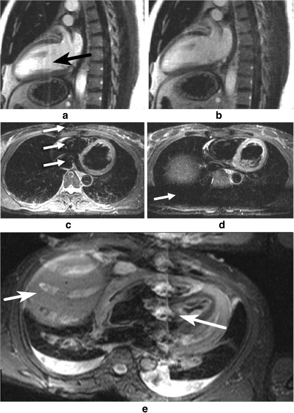
Figure 47.
CSF and pleural effusion ghosting. a-b) Long-axis LGE image: a) CSF ghosting is visible in the heart (arrow), b) CSF ghosting suppressed by a saturation band placed over the spinal canal. In this example the spatial location of the saturation is not very clear, this is presumably because of the timing of the spatial saturation that cannot immediately precede the imaging k-space centre, and the Mz recovery of other tissues due to gadolinium shortened T1. However, the T1 of CSF remains very long, which means that this stays well saturated. c-d) STIR transverse image: c) CSF ghost visible in the RV (arrows), d) CSF ghosting supressed by saturation band placed over the spine (arrow). e) Ghosting of the fluid in the pleural layers (pleural effusion) (arrows) in part due to its long T1. Pulsatility effects may also be present, contributing to the ghosting.

