
Figure 2.
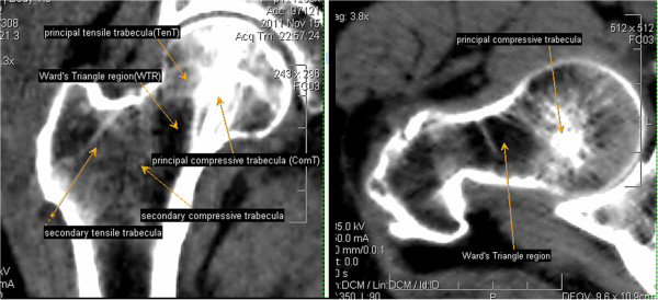
The location of the trabeculae and Ward's Triangle region internal proximal femur. (A) A mid-coronol image of proximal femur reveals: principal compressive trabecula; principal tensile trabecula; secondary compressive trabecula; secondary tensile trabecula and Ward’s Triangle region. (B) A transverse section of femoral neck reveals: principal compressive trabecula and Ward's Triangle region.