
Figure 5. Modelling the potential effect of D126N.
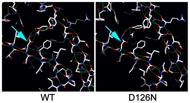
The human Nxt1 protein structure was used to model the Drosophila Nxt1 protein folding (WT) using Swisspdb viewer, and the region containing the mutated residue is shown. The side chain of D126 is indicated with an arrow. Computed H-bonds are shown in green. Running the model with the D126N substitution (arrow) revealed disruption of two computed H-bonds.