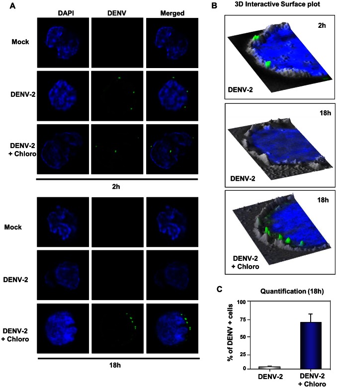
Figure 4. 3D microscopy of DENV-2 particles in purified plasmacytoid dendritic cells.
Freshly purified pDCs cultured with DENV-2 pre-treated or not with chloroquine (Chloro), or mock infected were stained with anti-DENV (green) and nucleus was colored with DAPI (blue). (A) pDC images (nucleus, virus and overlay) for mock, DENV-2 and chloroquine-treated plus DENV-2. Inhibition of endosomal acidification (chloroquine) allowed easier detection of DENV particles (DENV-2+Chloro at 2 h or 18 h stimulation). 2 h pDC incubation with DENV-2 was sufficient to detect viral proteins in contrast to the overnight (18 h) DENV-2-incubated pDCs when no virus was detected. (B) pDCs cultured with mock, DENV-2 or DENV-2+chloro were observed by 3D microscope. DENV staining (green) was merged with DAPI (blue)-colored nucleus and with phase contrast (grey). DENV particles were co-localized with pDC cell membrane at 2 h stimulation. Chloroquine allowed DENV-2 detection inside pDCs after 18 h of culture whereas DENV-2 alone did not. Panels shown microscopic images analyzed by 3D interactive surface plot. (C) Quantification of PDCs expressing DENV antigens without (DENV-2) and with chloroquine pre-treatment (DENV-2+Chloro) is shown as percentage of total analyzed cells.

