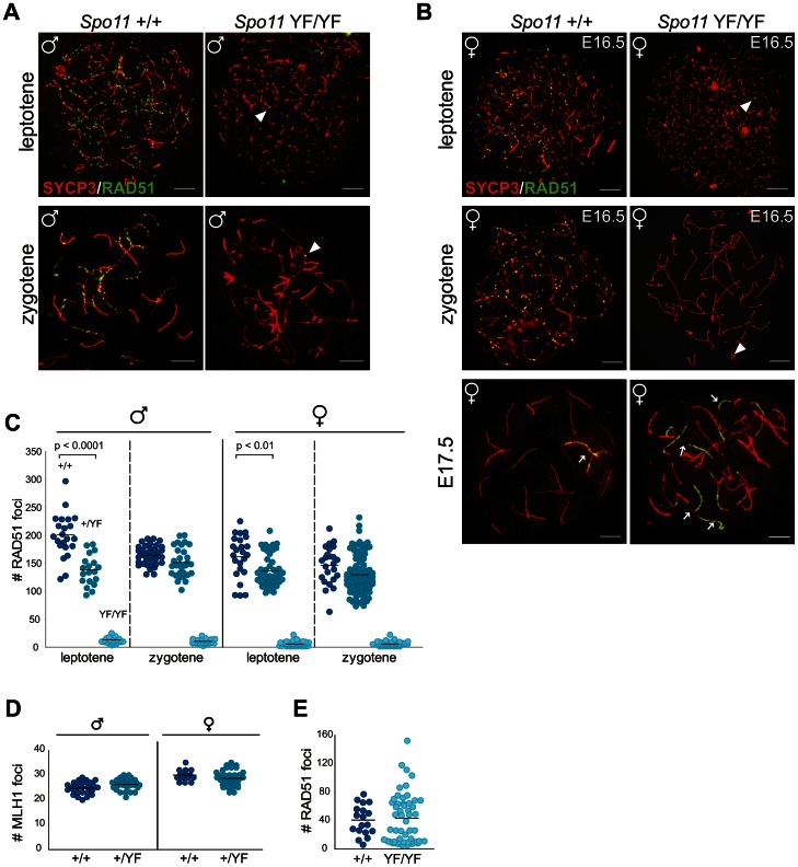
Figure 1. SPO11-dependent and -independent RAD51 foci in mouse meiocytes.
(A–C) The number of RAD51 foci decreases from leptotene to zygotene in Spo11+/+ and Spo11+/YF spermatocytes, whereas a few foci are detected in Spo11YF/YF spermatocytes and oocytes at both stages. (A–B) Double immunostaining with anti-SYCP3 (red), anti-RAD51 (green) of spermatocyte (A) and oocyte (B) nuclei from Spo11+/+ (A–B, left panel) and Spo11YF/YF (A–B, right panel) mice. Arrowheads point to RAD51 foci in Spo11YF/YF spermatocytes and oocytes, both leptotene and zygotene. Extensive accumulation of RAD51 along axial elements of one or few chromosomes (arrows) can be observed in both Spo11+/+ and Spo11YF/YF oocyte nuclei (B, lower panel). Size bars represent 10 µm. (C) The number of RAD51 foci was counted in Spo11+/+, Spo11+/YF, and Spo11YF/YF leptotene and zygotene spermatocytes and oocytes. Each dot represents the focus count of one nucleus. Black bars indicate mean number of foci. P values for the indicated comparisons (Mann-Whitney, two-tailed), and genotypes are indicated in the plot. (D) The number of MLH1 foci in pachytene spermatocyte nuclei was counted in Spo11+/+and Spo11+/YF mice. Black bars indicate the mean values. (E) Number of RAD51 foci at E17.5 in Spo11+/+ and Spo11YF/YF oocytes.

