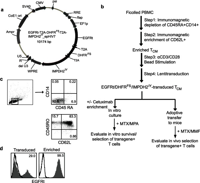
Figure 1. Experimental system for evaluating selection of gene-modified T cells in NSG mice.
(a), Plasmid map of construct containing drug resistance genes DHFRFS and IMPDH2IY, huEGFRt and T2A gene sequences (black) that was used to genetically alter primary human T cells. Lentiviral vector backbone (epHIV7) - related sequences are depicted in grey. (b), Schematic for isolation, genetic modification and selection of primary human T cells. (c), CD45RA and CD14 staining of mononuclear cells after sorting from PBMC (top), and CD62L and CD45RO staining of T cells after enriching from PBMC (bottom). The percent positive cells in each quadrant are indicated. (d), Primary human T cells were transduced with above lentiviral vector; transduced T cells and immunomagnetic sorted EGFRt+ T cells were stained for EGFRt expression and analyzed for transduction efficiency by flow cytometry. The percent EGFRt+ T (grey histogram; isotype control-dark line) cells are depicted.

