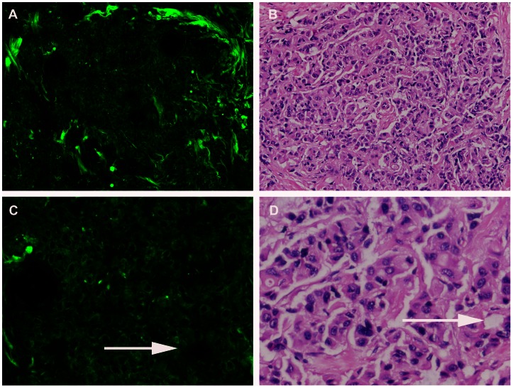
Figure 5. Breast cancer.
Multiphoton microscopy (MPM) image (A) and corresponding hematoxylin-eosin (H-E) image (B) show cancer cells displayed marked cellular and nuclear pleomorphism. Cancer cells are characterized by irregular size and shape, enlarged nuclei, and increased nuclear-cytoplasmic ratio. Glandular structure consisting of tumor cells is evident (as indicated by arrow) shown in C, D). (Original magnifications 63x [A]; 20x [B]).

