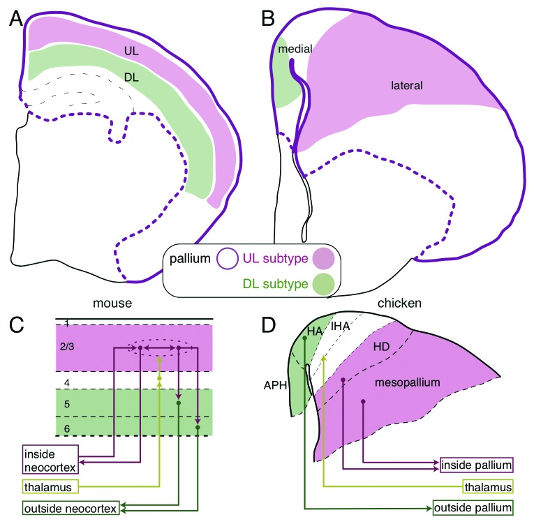
Figure 1. Comparison of the mammalian and avian pallial architectures. (A) The layered neocortex of the mouse. The upper and deep layer (UL and DL) neuron subtypes are tangentially arranged in the pallium. (B) UL and DL neuron subtypes are arranged separately in the medial and lateral domains of the chick pallium, respectively. (C and D) Similarity of the neural circuits in the mammalian neocortex and the avian pallium. (C) The columnar neural circuit in the mammalian neocortex. The input from the thalamus terminates in the layer 4. The information is transferred to and processed in the UL (layer 2/3) neurons that are connected with each other inside the neocortex, and finally output by the DL (layer 5 and 6) neurons to extracortical targets. (D) In the avian pallium, the thalamic input is received by the neurons in the central domain of the hyperpallium (IHA), which is sandwiched by the medial (HA and APH) and lateral domains (HD and mesopallium). The medial and lateral domains project to the intrapallial and extrapallial targets, respectively.

An official website of the United States government
Here's how you know
Official websites use .gov
A
.gov website belongs to an official
government organization in the United States.
Secure .gov websites use HTTPS
A lock (
) or https:// means you've safely
connected to the .gov website. Share sensitive
information only on official, secure websites.
