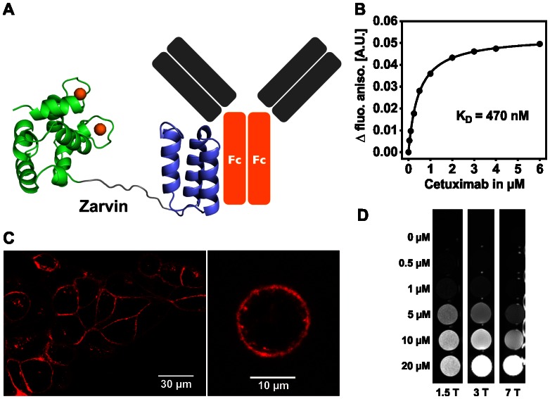
Figure 1.
Binding properties and relaxometric properties of Zarvin. (A) Cartoon representation of Zarvin bound to the Fc part of an IgG antibody. Two calcium ions (spheres) are bound to Parvalbumin (green), which is connected with the Z domain (violet) via a decaglycine linker (grey). (B) Fluorescence anisotropy titration experiment. Increasing amounts of the monoclonal IgG antibody Cetuximab were added to a 100 nM concentration of Zarvin-Atto-465. (C) Confocal microscopic analysis of the complex Cetuximab:Zarvin-D72C-Atto 594 binding to the EGF receptor located in the cell membrane of A431 cells. Left, cell assembly; right, single cell; control experiments (Figure S4) (D) Relaxometric properties of Zarvin:(Gd3+)2 at three different field strengths employing an inversion recovery TSE experiment. A diluted solution of rising concentrations of Zarvin:(Gd3+)2 was investigated to find the limiting concentration which still produces a visible contrast towards the buffer control (0 µM). The picture is displayed with an inversion time TI which zeroes the signal of the buffer control (appears black).

