
Figure 2. Facial growth remodelling maps.
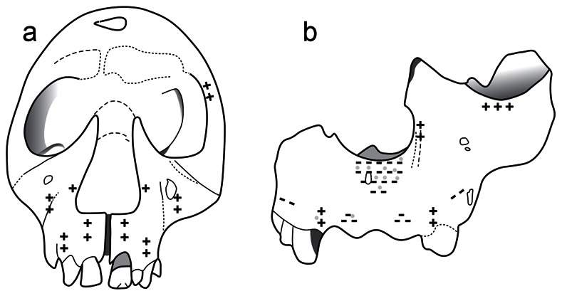
(A) Facial growth remodelling of the H. erectus specimen KNM-WT 15000 from Kenya, dating from ∼1.5 my showing depository fields (+) over most aspects of the anteriorly facing maxilla. Taphonomic alterations prevented a more complete analysis of the periosteal surface of this specimen which was only studied by SEM. (B) Facial growth remodelling of the specimen ATD6-69 representing H. antecessor, the oldest known European hominin species dating to 900–800 ky. SEM and confocal microscopy data showed resorptive fields (−) throughout the naso-alveolar clivus of this hominin, a characteristic shared with H. sapiens. Gray circles indicate the areas spot-mapped using the portable confocal microscope (PCSOM).