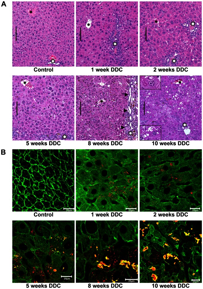
Figure 1. Pathomorphological changes in mouse liver tissue during DDC-feeding.
A: Hematoxylin-eosin staining. Pathomorphological changes in mouse liver tissue (white asterisks: portal tracts; black asterisks: central veins; black arrows: fibrosis and ductular reaction). During early stages (1, 2 and 5 weeks) of DDC-feeding, hepatocytes increase in size compared to control. Mild and focal inflammation was observed in the lobular parenchyma. Eight and 10 weeks of DDC-feeding shows additional occurrence of steatosis, hepatocellular ballooning and MDBs (enlarged inset left upper corner: ballooned hepatocyte containing an MDB; left lower corner: hepatocyte with steatosis). B: Double immunofluorescence (red: p62; green: keratin 8/18). In controls only the keratin cytoskeleton network (green) is visible in the hepatocytes, during DDC-intoxication (1–2 weeks) pericellular fibrosis is recognizable; at 5 weeks p62-containing aggregates appear which develop to MDBs, containing p62 and keratin 8/18 (green+red→yellow) at weeks 8 and 10. All scale bars are 20 µm.

