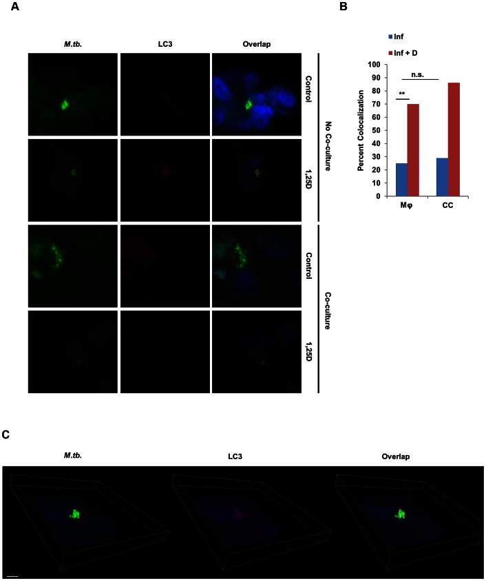
Figure 7. Co-culture with SAECs does not enhance autophagy in Mtb-infected macrophages.
(A) Representative bright-field microscopy of infected macrophages cultured in the absence or presence of 1,25D and/or a transwell containing SAECs. Cells were fixed and probed with an antibody against Mtb, LC3, and the nuclear stain DAPI. (B) Percent of Mtb that colocalized with LC3 signal under each condition as determined by counting populations of infected macrophages across at least 10 confocal images, with at least two infected cells per image. Statistical significance was determined by a two-tailed Fisher's exact test (**P<0.01). (C) 3-dimensional rendering of confocal stacks of infected macrophages treated with 1,25D to demonstrate colocalization of LC3 and Mtb signal.

