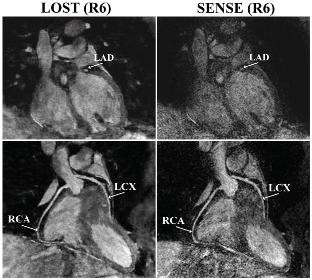
Figure 2.
An example coronal slice (top) and reformatted coronal image (bottom) of a subject using B1-weighted LOST with 6-fold random undersampling (left) and SENSE with 6-fold uniform undersampling. A cross-section of the LAD is visualized clearly with the proposed technique, whereas noise amplification is apparent in the SENSE-reconstructed image (RCA: right coronary artery, LAD: left anterior descending, LCX: left circumflex).

