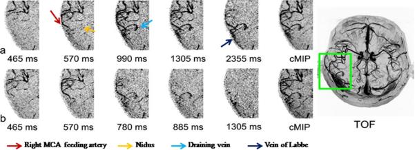
Figure 9.

Representative temporal phases of axial dMRA MIP images and collapsed MIP image (cMIP) acquired using 4-bolus TrueSTAR (a) and standard TrueSTAR (b) from the AVM patient. TOF MRA MIP image after the injection of gadolinium contrast agent is shown on the right side. Multi-bolus dMRA provided improved visualization of the nidus and draining veins.
