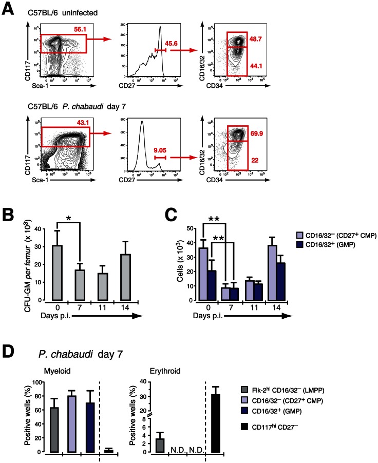
Figure 3. Effect of acute infection with P. chabaudi on myeloid progenitor cells in the bone marrow.
Infection of C57BL/6 female mice (6–8 weeks) with P. chabaudi resulted in a transient decrease of myeloid lineage progenitors in the bone marrow (BM) during acute infection. A. BM cells from uninfected controls and malaria-infected animals at day 7 of infection with P. chabaudi were co-stained with antibodies against lineage markers, Sca-1, CD117 (c-Kit), CD27, CD34, CD16/32 (FcγRII/III) and separated as indicated. Doublets were electronically excluded prior to analysis. Data are representative for 19 infection experiments with numbers in individual plots indicating the frequency (in %) of the respective population. B. Analysis of granulocyte-monocyte colony forming units in semi-solid media from total BM during acute malaria. Data are from two experiments with 4–5 BM samples per experiment and shown as mean ± SEM of total number of CFU-GM per femur (*: P≤0.05, two-tailed Student's t-test). C. Absolute cell numbers of the CD27+ CMP (light blue bars) and the GMP (blue bars) progenitor populations during the course of a malaria infection. Data represent mean ± SEM of cell number per femur/pair obtained from 19 infection experiments with 4–5 mice per group (**: P≤0.01, Mann-Whitney U-test). D. Single cell in vitro cultures that support myeloid or erythroid differentiation of FACS purified hematopoietic BM progenitors obtained at day 7 after infection with P. chabaudi. Data are shown as mean ± SEM of individual positive wells (in %) of three infection experiments with 5 mice per subset (N.D. = not detected).

